Many different factors and conditions can cause CKD including:
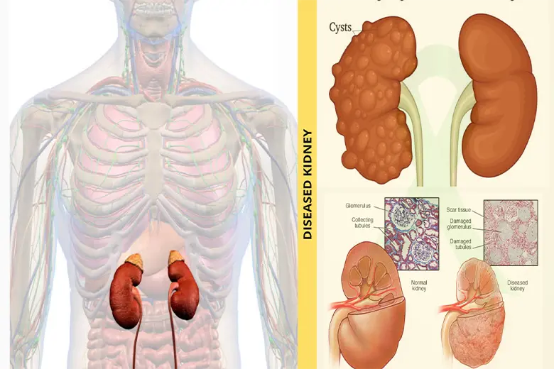
Polycystic kidney disease
Glomerulonephritis
Lupus
Obstructive uropathy
Reflux nephropathy
Reflux nephropathy
Hypoplastic kidneys
Hypertension
Diabetes etc.
Chronic Kidney Disease (CKD) // end stage renal failure that has no cure and requires renal replacement therapy, that is, dialysis or kidney transplant.
Mesenchymal stem cell has therapeutic potential in improving renal function deficits, and healing tubular injury. Mesenchymal stem cells can integrate into renal tubular cells and can differentiate into mesangial cells. Moreover, these cells stimulate the kidney’s own stem cells (resident stem cells) and release growth factors which promote the survival of renal cells, thus, initiating a natural recovery.
How It Is Done
Thorough evaluation with ultrasonography /CT scan and complete haematological and biochemical tests including end stage kidney scoring is done.
Under local anaesthesia 100 cc fat is aspirated. If possible approximately 150 cc of bone marrow is also aspirated from iliac crest or Tibia.
Fat and bone marrow are processed to isolate various types of stem cells (haematopoietic, mesenchymal, endothelial and very small embryonic stem cells).
In cases which are very compromised allogenic umbilical cord derived mesenchymal stem cells is an alternative option.
These cells are injected
Intravenous
Into Renal artery via angiographic guidance Intravenous route needs multiple administration and higher dose of cells
What To Expect
80 % patients showed significant improvements in Kidney function Tests.
Improvements started to be observed as early as by 4 weeks.
Well maintained up to 9 months.
Creatinine from average baseline of 148.6 μmol/L, to 120.2 μmol/L.
UREA: from average baseline of 34.71 mmol/L, to 14.40mmol/L.
Dialysis was reduced to 1-2 times a week from 4 times a week in 40 % of patients.
GFR increased to the range of 5-12 mL/min/1.73 m2 in 60 % of patients.
Color Doppler ultrasound showed size of both sides of kidney slightly increased with visible adequate blood flow in the both kidneys in 40% of patients.
Albumin and WBC in urine reduced to within normal limits by 4 months in 75 % of patients.
Blood pressure was also well controlled and remained stable in 75 % patients.
Associated Type 2 diabetics achieved better blood sugar control along with renal improvement.